Welcome to the new updated XLHLink.com!
REsources

resources

4-year-old female with spontaneous XLH*

Case summary

Findings/outcomes

  • The patient was seen by multiple specialists and after referral to an endocrinologist; the patient's case was subsequently followed by a nephrologist
  • This patient’s disease symptoms continued to progress despite therapy with oral calcitriol and potassium phosphate

*The information for this case study was provided courtesy of Dr. Anthony Portale, Director of the Pediatric Dialysis Program, UCSF Benioff Children’s Hospital, San Francisco. This case study represents a real patient and is intended to be illustrative, not a recommendation for treatment or management. This case study does not claim to represent typical results.

The letter i within a diamond denoting a callout specific to the XLH disease-monitoring program

Although X-linked hypophosphatemia (XLH) is primarily an inherited disease, 20% to 30% of cases arise spontaneously.1,2

Medical history

18-19 months: Pediatric evaluation

  • 18-month-old female presented with leg bowing, wide-based gait, delayed walking, and poor growth; fear of climbing stairs
  • X-rays: indicative of metaphyseal dysplasia
  • Other clinical findings: strabismus, suspected to contribute to walking delay; underwent strabismus surgery

28 months: Orthopedic evaluation

  • Physical exam: short stature, height less than 5th percentile since age 15 months (see Growth chart); worsened bowing and waddling gait
  • X-rays: significant bilateral genu varum with mild beaking and sloping of the proximal tibial metaphysis; metaphyseal-diaphyseal angles of bilateral proximal tibiae measured 16 degrees; bilateral irregularity of the distal femoral metaphysis also noted; femoral heads were normally located
  • Differential diagnosis: possible Blount disease, metaphyseal dysplasia, or physiologic genu varum

29 months: Genetics evaluation

  • No family history of bone disease
  • Suspected diagnosis: metaphyseal dysplasia
  • Underwent genetic testing for Schmid metaphyseal chondrodysplasia and Shwachman-Diamond syndrome; results were negative for pathologic variants
  • Diagnosis: unknown, referred to skeletal dysplasia clinic

31 months: Endocrinology evaluation

  • Physical exam: bilateral genu varum
  • X-rays: bilateral mild coxa vara, tibial varus (left greater than right) with metaphyseal irregularity and fraying of the distal femur, tibia, and ulna; metaphyseal widening at wrists
  • See Laboratory test results below
  • Other findings: 2nd percentile for weight; 1st percentile for height (see Growth chart below)
Growth chart: Stature for age percentiles, girls aged 2-6 years3†

The reference percentiles on the graph are combined from the 2 clinical growth charts for females 2-20 years of age provided by the Centers for Disease Control and Prevention.

History of growth progression:

  • Patient had short stature
  • At 31 months, the patient was in the 1st percentile for height

Diagnosis and initial treatment

  • Hypophosphatemic rickets; positive for PHEX mutation; negative in parents—spontaneous X-linked hypophosphatemia (XLH)
  • Treatment: oral calcitriol and potassium phosphate
The letter i within a diamond denoting a callout

For patients with XLH, the diagnosis process can be long and laborious, and proper specialist referral can be key to early disease management.

Disease progression

39 months: Nephrology follow-up evaluation

  • Patient continued to take oral calcitriol and potassium phosphate
  • Physical exam: wide-based gait; mild genu varum; intercondylar distance 1.0 cm; bilateral mild tibial bowing
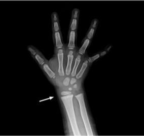
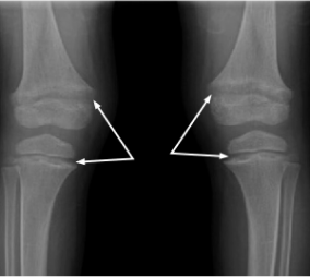
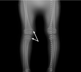
  • X-rays: mild metaphyseal fraying at distal radius and ulna; metaphyseal widening and fraying at distal femur and proximal tibia (see X-rays 1-3)

54 months

  • Further management options explored due to continued disease progression
  • See Laboratory test results below
X-ray 1: Right wrist
X-ray of right wrist

54 months

  • Further management options explored due to continued disease progression
  • See Laboratory test results below
X-ray 2: Knees
X-ray of knees
X-ray 3: Legs
X-ray of legs
54 months: mild metaphyseal fraying at the bilateral distal radius and ulna, and to a lesser extent of the bilateral distal femoral metaphyses.

Laboratory results

Test (reference range unit) 31 months (results) 54 months (results)

Serum phosphorus

(4.3-6.8 mg/dL)
2.9 2.5
TmP/GFR (4.0-5.2 mg/dL) 2.4 n/a
25(OH)D (≥20 ng/mL) 41 31
ALP (<156-369 U/L) 461 404
Serum calcium (9.2-10.5 mg/dL) 9.4 9.9
PTH (<10-65 pg/mL) 57 31

Indicates normal range, age, and sex matched. Note that normal range values may vary depending on reference dataset. The ranges in this table were provided by the treating physician. Colored values are outside of the normal range provided by the physician and can raise suspicion of XLH.

25(OH)D=25-hydroxyvitamin D; ALP=alkaline phosphatase; PTH=parathyroid hormone; TmP/GFR=ratio of tubular maximum reabsorption of phosphorus to glomerular filtration rate.

The letter i within a diamond denoting a callout

This patient’s disease symptoms continued to progress despite therapy with oral calcitriol and potassium phosphate.

15-year-old female with spontaneous XLH§

Case summary

Findings/outcomes

  • The goal of management aimed to optimize bone health before growth plate closure
  • Regardless of provider, asking about mental health was an important element of care
  • Once growth potential was reached, management of XLH was not over; goals shifted from promoting bone growth to fracture prevention and maximizing musculoskeletal health
§The information for this case study was provided courtesy of Dr. Anthony Portale, Director of the Pediatric Dialysis Program, UCSF Benioff Children’s Hospital, San Francisco. This case study represents a real patient and is intended to be illustrative, not a recommendation for treatment or management. This case study does not claim to represent typical results.
The letter i within a diamond denoting a callout

Symptoms of XLH are nonspecific when considered individually and vary between patients. Specialist referral is key to early disease management.

Medical history

Birth

  • Full-term infant with no abnormalities detected

12 months

  • Signs consistent with pain upon weight bearing; limping; bowed legs at 18 months

Family history

  • Unremarkable

30 months

  • X-rays: bilateral bowing with irregular and widened metaphyses at tibia and femur

34 months: Key fasting laboratory findings (reference range)

  • Serum phosphorus: 2.4 mg/dL (4.3-6.8 mg/dL)
  • 25-hydroxyvitamin D: 39 ng/mL (≥20 ng/mL)
  • Alkaline phosphatase: 512 U/L (156-369 U/L)
  • Serum calcium: 9.2 mg/dL (9.2-10.5 mg/dL)
  • Parathyroid hormone: 85 ng/L (10-65 ng/L)
Growth chart: Stature for age percentiles, females aged 2-20 years

||Indicates normal range, age, and sex matched. Note that normal range values may vary depending on reference dataset. These ranges were provided by the treating physician. Colored values are outside of the normal range provided by the physician and can raise suspicion of XLH.

The reference percentiles on the graph are combined from the 2 clinical growth charts for girls 2-20 years of age provided by the Centers for Disease Control and Prevention.

History of growth progression:

  • Patient had short stature. Linear growth accelerated from 8 to 10 years of age, consistent with early growth spurt

Diagnosis and initial treatment

  • Spontaneous XLH
  • Treatment: oral calcitriol and phosphate

Disease progression

1-3 years of age: Managed under orthopedic care

  • Optimal control of metabolic bone disease difficult to achieve
  • Patient had short stature (see Growth chart above)

3 years of age: Referred to endocrinology

5.9 years of age
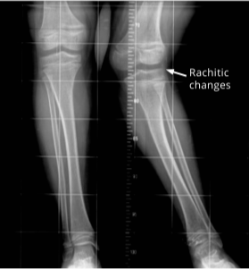
  • Musculoskeletal presentation with limb deformity in femur, tibia, and fibula; rachitic changes (see X-ray 1)
X-ray 1: Limb deformity
X-ray of legs indicating limb deformity
5.9 years of age: limb deformity: femur, tibia, and fibula; rachitic changes; lateral angulation at left knee.
The letter i within a diamond denoting a callout

Although this patient’s laboratory results improved, there was no height velocity increase. At 3 years of age, it was concluded that biochemical optimization was not met; patient was then seen monthly to tailor therapy.

7 years of age: Metabolic bone disease under control

  • Surgery of lower left extremity discussed, postponement recommended
  • Reported some classmates teased her for wearing a brace
  • Parents reported good activity level

3-9 years of age

  • Growth velocity normal (see Growth chart above)

9.5 years of age

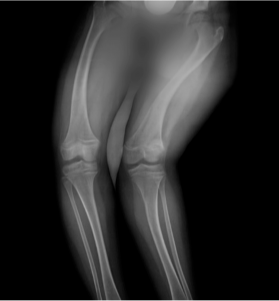
  • Musculoskeletal presentation at prominent genu valgum, severe angulation at left knee with knees touching (see X-ray 2)
X-ray 2: Severe left knee lateral angulation
X-ray of legs indicating severe left knee lateral angulation
9.5 years of age: prominent genu valgum of left lower extremity; severe lateral angulation at left knee, with knees touching; 16 cm between medial malleoli, minimal medial angulation of right lower extremity; wide-based gait.
The letter i within a diamond denoting a callout

As this patient matured, awareness of disability and obvious orthopedic deformities increased. Regardless of provider, asking about her mental health was an important element of care.

10 years of age
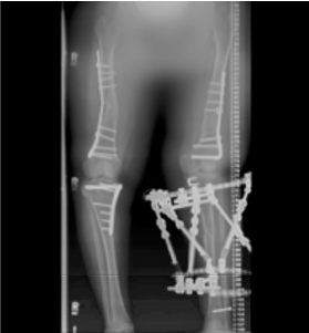
  • Linear growth accelerated consistent with early growth spurt; patient was at the 5th percentile for height (see Growth chart above)
  • Underwent bilateral epiphyseal arrest surgery (epiphysiodesis) (see X-ray 3)
  • Increasing complaints of muscle weakness and left hip, leg, and back pain
  • Tired very easily and could walk only short distances without resting
  • Limb deformity worsened despite treatment with oral calcitriol and phosphate
    • Exhibited signs of secondary varus of right knee as in windswept deformity
X-ray 3: Windswept deformity
X-ray of legs indicating severe left knee lateral angulation
10 years of age: epiphyseal arrest surgery; windswept deformity.

Between 12.5 and 14.5 years of age

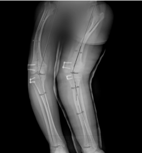
  • Patient underwent successful osteotomies of femurs and left tibia (see X-ray 4)
  • Homeschooled
X-ray 4: Osteotomies
X-ray of osteotomies of femurs and left tibia
12.5 to 14.5 years of age: osteotomies of femurs and left tibia.
The letter i within a diamond denoting a callout

Once growth potential was reached, XLH management continued, including monitoring of biochemical bone health to promote healing and peak bone mass. As the patient aged, her disease management transitioned from promoting bone growth to fracture prevention and maximizing musculoskeletal health.

49-year-old male with hereditary XLH#

Case summary

Findings/outcomes

  • The patient was not accurately diagnosed with XLH until age 44
    • There is a spectrum of disease for XLH,4 and the patient’s symptoms were not previously attributed to XLH
  • Symptoms of XLH progressed throughout adulthood and had long-term impact. The patient:
    • Required surgeries and developed worsening pain
    • Required the use of assistive walking devices
    • Developed hearing loss
    • Needed dentures
  • Alternative therapeutic options were explored

#The information for this case study was provided courtesy of Dr. Kathryn McCrystal Dahir, Professor of Medicine, Department of Endocrinology, Vanderbilt University Medical Center, Nashville. This case study represents a real patient and is intended to be illustrative, not a recommendation for treatment or management. Results may vary. This case study does not claim to represent typical results.

The letter i within a diamond denoting a callout

It is important to diagnose XLH accurately and early, as the progressive nature of XLH leaves patients susceptible to short stature, fractures, limited function, and pain.1,5,6

Medical history

During childhood

  • 6 years of age: humerus fracture from jumping off a moving truck
  • Poor dentition of both primary and secondary teeth with multiple cavities and abscesses
  • 11 years of age: reported “bone disease” due to chronic bone and joint pain
    • Pediatrician diagnosed post-streptococcal arthritis
  • Mild leg bowing; reported short stature compared to peers

Early to mid-20s

  • Lost most teeth by early 20s
  • Acute worsening of bone and joint pain
    • Suspected inflammatory arthritis; treated with anti-inflammatory medication

44 years of age

  • Presented with acute exacerbation of chronic weakness and right hip pain
    • Required the use of prescription pain medication, including hydrocodone acetaminophen
  • Reported no previous diagnosis of rickets or any medical management of low phosphorus

Family history

  • Short stature and bowing of the lower legs in mother (height, 4’11”), 3 brothers, and 2 nieces; all had multiple fractures

Physical exam

  • Height, 5’3”; weight, 230 lbs
  • Ambulating with assistance of a walker; knees in varus alignment
  • Pain with internal rotation of hips (left worse than right); 4/5 motor strength in both proximal and distal muscle groups
  • Poor dentition and missing most secondary teeth

X-rays

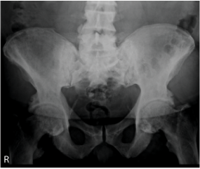
  • Pelvis and hips: diffuse increased density of the bones, particularly in the iliac wings and lumbosacral spine; enthesopathic calcifications extending from the superior acetabular walls bilaterally (see X-ray 1 below); right subtrochanteric pseudofracture identified, which was incomplete along the medial cortical region (see X-ray 2 below)
  • Femurs: pseudofractures in the subtrochanteric right femur and mid-left femoral diaphysis; healing stress fracture present in the medial cortex of the left femoral neck; enthesophytes and bilateral hip osteoporosis also noted (see X-ray 3 below)
  • See Laboratory test results below
X-ray indicating disease progression in pelvis and hips
X-ray 1: Disease progression in pelvis and hips

44 years of age: diffuse increased density of iliac wings and lumbosacral spine; bilateral enthesopathic calcifications extending from the superior acetabular walls.
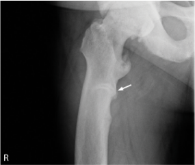
X-ray of subtrochanteric pseudofracture
X-ray 2: Subtrochanteric pseudofracture

44 years of age: right subtrochanteric pseudofracture (arrow) incomplete along the medial cortical region.
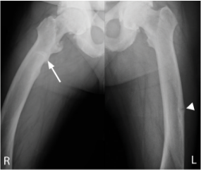
X-ray of pseudofractures in femurs
X-ray 3: Pseudofractures in femurs

44 years of age: pseudofractures in the subtrochanteric right femur (arrow) and mid-left femoral diaphysis (arrow head); healing stress fracture present in left femoral neck medial cortex; enthesophytes and bilateral hip osteoporosis.

The letter i within a diamond denoting a callout

The patient exhibited XLH signs and symptoms throughout childhood and young adulthood, and was not diagnosed until his mid-40s.

Diagnosis and initial treatment

  • Hereditary XLH
  • Treatment: calcitriol and phosphate supplementation

Disease progression (post-treatment)

44 years of age

After XLH diagnosis, underwent left hip replacement surgery

  • Developed worsening right hip pain; underwent right hip replacement surgery 3 months later

46 years of age

Developed worsening left shoulder and knee pain requiring multiple orthopedic visits for steroid injections

  • Developed loosening of hardware in left hip due to osteomalacia and continued to require frequent steroid therapy
  • Reported being compliant to conventional therapy

47 years of age

Underwent knee replacement therapy evaluation

  • Unable to work and required disability benefits
  • Dependent on assistive walking devices
  • Developed ringing in ears and hearing loss
  • Referred to pain management; nerve pain medication added to regimen with little pain improvement
  • See Laboratory results below

49 years of age

Underwent left knee replacement; advised a right total knee replacement was needed as well

  • Insurance started denying coverage for calcitriol and phosphate supplementation on the basis that these medications are supplements/vitamins
  • Medication was continued with coverage through a grant
Patient reported compliance with conventional therapy; developed worsening back, neck, and hand pain
Could not afford dentures and had difficulty maintaining adequate nutrition

3 months later: Alternate therapeutic options were explored

The letter i within a diamond denoting a callout

The patient initiated oral phosphate and active vitamin D and was compliant. Symptoms of XLH continued to progress, leading to a search for alternative treatment options.

Laboratory results

Test (reference range** unit) 44 Years (results) 47 Years (results)

Serum phosphorus

(2.4-5.7 mg/dL)
2.1 2.1
1,25(OH)2D (20-80 ng/mL) 31 25
25(OH)D (25-80 ng/mL) 33 37
BSAP (6.5-20.1 mcg/L) 39.2 32
PTH (16-77 pg/mL) 95 48
Creatinine (0.72-1.25 mg/dL) 0.83 1.12
FGF23 (<180 RU/mL) 73 383

**Indicates normal range, age, and sex matched. Note that normal range values may vary depending on reference dataset. These ranges were provided by the treating physician. Colored values are outside of the normal range provided by the physician and can raise suspicion of XLH.

1,25(OH)2D=1,25-dihydroxyvitamin D; 25(OH)D=25-hydroxyvitamin D (calcifediol); BSAP=bone-specific alkaline phosphatase, also known as BAP; FGF23=fibroblast growth factor 23; PTH=parathyroid hormone.


Connect the symptoms and take the next step

Icon of an eye

Suspect XLH?

Connect their symptoms and accurately diagnose with the right tests.

Icon of a microscope

Diagnosing XLH

Learn more about how to accurately diagnose XLH through testing.

Icon of a gear

Managing XLH

Manage XLH early to prevent progressive bone damage.

Stay connected

Set up time with a representative to talk more about XLH, or sign up for news and information about XLH.

References:

1. Dahir K, Roberts MS, Krolczyk S, Simmons JH. X-linked hypophosphatemia: a new era in management. J Endocr Soc. 2020;4(12):bvaa151. doi:10.1210/jendso/bvaa15 2. Gaucher C, Walrant-Debray O, Nguyen TM, Esterle L, Garabédian M, Jehan F. PHEX analysis in 118 pedigrees reveals new genetic clues in hypophosphatemic rickets. Hum Genet. 2009;125(4):401-411. doi:10.1007/s00439-009-0631-z 3. Centers for Disease Control and Prevention. Clinical growth charts. 2 to 20 years: girls. Published May 30, 2000. Accessed May 21, 2019. https://www.cdc.gov/growthcharts/data/set2clinical/cj41c072.pdf 4. Ruppe MD. X-linked hypophosphatemia. In: Adam MP, Everman DB, Mirzaa GM, et al., eds. GeneReviews®. Seattle (WA): University of Washington, Seattle; February 9, 2012. Updated April 13, 2017. https://www.ncbi.nlm.nih.gov/books/NBK83985/ 5. Carpenter TO, Imel EA, Holm IA, Jan de Beur SM, Insogna KL. A clinician’s guide to X-linked hypophosphatemia. J Bone Miner Res. 2011;26(7):1381-1388. doi:10.1002/jbmr.340 6. Hamilton AA, Faitos S, Jones G, Kinsley A, Gupta RN, Lewiecki EM. Whole body, whole life, whole family: patients’ perspectives on X-linked hypophosphatemia. J Endocr Soc. 2022;6(8):bvac086. doi:10.1210/jendso/bvac086